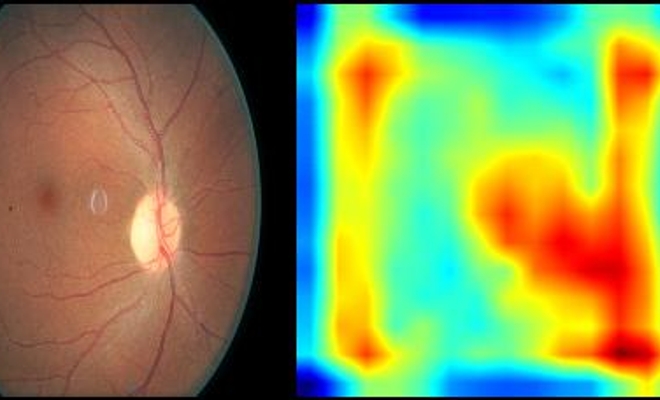
VISION-DR: Visual Insights and Saliency Integrated Overlay Neural Network for Diabetic Retinopathy

VISION-DR: Visual Insights and Saliency Integrated Overlay Neural Network for Diabetic Retinopathy
Overview
Welcome to the VISION-DR project, a groundbreaking computer vision approach for the diagnosis of diabetic retinopathy (DR). This project leverages a VGG-based convolutional neural network (CNN) trained on fundus camera images to classify the severity of DR and generate saliency maps for enhanced interpretability. Our tool is designed to assist healthcare professionals by providing insights into the model’s decision-making process, ultimately supporting more accurate and informed clinical judgments.
Project Description
Diabetic retinopathy is a leading cause of vision impairment among diabetic patients. Early detection is crucial for effective management and treatment. VISION-DR aims to automate the detection and classification of DR into various stages of severity using deep learning techniques, while also enhancing transparency and trust through the use of saliency maps.
Key Features
- VGG-16 Based Model: Utilizes the robust feature extraction capabilities of the VGG-16 architecture to classify the severity of DR.
- Saliency Maps: Generates visual explanations of the model's predictions, highlighting the specific areas within the images that influence the classification decisions.
- Clinician Support: Designed to support, not replace, the clinical judgment of physicians by providing additional insights and focusing attention on areas of potential concern.
Methodology
Dataset
The model is trained on the Kaggle Diabetic Retinopathy Detection Dataset, which includes over 35,000 high-resolution retina images labeled by clinicians.
Model Architecture
The VGG-16 architecture, pre-trained on ImageNet, is adapted for this task by unfreezing all layers and modifying the classifier to include:
- A linear layer
- ReLU activation function
- Dropout layer (0.5 rate)
- Sigmoid activation for binary classification
Training Process
- Loss Function: Cross-entropy loss
- Optimizer: Adam optimizer with a learning rate of 0.001
- Evaluation Metric: Area Under the Receiver Operating Characteristic Curve (AUC-ROC) due to class imbalance
Interpretability
We employ Gradient-weighted Class Activation Mapping (Grad-CAM) to produce saliency maps. These maps highlight the regions in the input images that are most important for the model's predictions, aiding in the verification and understanding of the model's decisions.
Results
The model achieved robust performance with an AUC-ROC score plateauing near 0.9, indicating a high probability of accurately distinguishing between different severity levels of DR. Saliency maps provided visual confirmation of the regions influencing the model's decisions, demonstrating the model's capability to identify clinically relevant features.
Future Work
Image Segmentation
Developing an image segmentation system to delineate detailed structures within fundus images could further enhance diagnostic accuracy.
Longitudinal Analysis
Analyzing sequential fundus images of the same patients to track the progression of DR over time can lead to more personalized treatment decisions.
Integration of Medical Knowledge
Incorporating established medical knowledge into the training process through regularization techniques to improve model reliability and generalization.
Conclusion
VISION-DR demonstrates the potential of computer vision models to enhance the diagnosis of diabetic retinopathy. By providing accurate classifications and interpretability through saliency maps, this tool supports healthcare providers in making more informed decisions, ultimately contributing to better patient outcomes. As we continue to refine and expand this project, the integration of AI in medical diagnostics becomes increasingly feasible and beneficial.
References
For a detailed list of references, please refer to the complete research paper.


